The nematode, Enterobius vermicularis (pinworm), is the most common helminthic infection in North America, and are a common incidental finding on appendectomy. Pinworms typically infect school-age children via the fecal-oral route, however inhalation of eggs can occur.
Grossly, pinworms may be visible as small (2 - 13 mm), ivory-white worms with long, pointed tails. Microscopically on cross-section they have a thick cuticle, lateral alae (wings), and visible organs which may include intestines, and ovaries/testes. Eggs may also be seen within female worms or in isolation. They may be differentiated from vegetable matter as their exterior and cuticle does not stain for PAS/PASD.
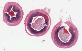
This slide shows H&E stained cross sections of the appendix. See Related Content for PASD stain and proximal and distal sections.
Sidiropoulos, K.G., Ngan, B. Appendix, Enterobius vermicularis, H&E stain. Digital Laboratory Medicine Library, Dept of Laboratory Medicine & Pathobiology, University of Toronto. Published
. Accessed December 17, 2025. https://dev.dlml.cflabs.ca/image/appendix-enterobius-vermicularis-he-stain-lmp19163