The patient presented with a 36 hour history of nausea and perimubilical pain which migrated to McBurney's point. U/S demonstrated a distended appendix with a dumbbell shape, consistent with acute appendicitis. The patient underwent an appendectomy where a neoplasm was noted arising from the tip of the appendix and extending into the periappendiceal tissue.
Case Discussion
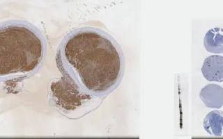
Well-differentiated neuroendocrine tumours are low-grade tumours with favourable prognosis, but should be condidered of uncertain malignant potential. They are found in approximately 1.5% of patients undergoing appendectomy. They are composed of small, polygonal cells forming nests and cords with monotonous nuclei, inconspicuous nucleoli and a characteristic stippled or "salt and pepper" chromatin. The tumour cells may be functional and can produce hormones, most commonly serononin, which can be seen as small eosinophilic granules in the cytoplasm of the tumour cells. Immunohistochemistry demonstrates that the tumour cells stain positively for serotonin and neuroendocrine markers (chromogranin and synaptophysin). Further surgical management (i.e. right hemi-colectomy) may be necessary if the tumour is greater than 2 cm and/or extends beyond the appendix (as in this case). This is a chromogranin stain.
Forse, C., Chang, M. Appendix, well-differentiated neuroendocrine tumour, chromogranin. Digital Laboratory Medicine Library, Dept of Laboratory Medicine & Pathobiology, University of Toronto. Published
. Accessed December 17, 2025. https://dev.dlml.cflabs.ca/image/appendix-well-differentiated-neuroendocrine-tumour-chromogranin-lmp40251