13 month old male infant with a tumour on the shoulder.
Case Discussion
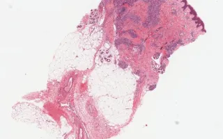
Tufted angiomas are benign vascular lesions which often occur on the neck or shoulders of children or young adults. Microscopically, they are comprised of nodules of tightly packed capillaries which may involve the dermis and superficial subcutis. Crescentic cleft-like vessels or semilunar lymphatic channels are generally seen at the periphery of some of the capillary tufts (nodules). These vessels are lined by unremarkable endothelial cells (CD31 positive) and surrounded by pericytes (smooth muscle actin positive). D2-40, a marker of lymphatic endothelial cells, is expressed in the crescentic cleft like vessels, but the proliferative capillaries are negative. The capillary lumina are not readily evident in many areas. GLUT-1 was negative in this case; it is positive in juvenile hemangiomas.
This slide shows H&E stain, see Related Content for CD31, D2-40, and SMA stains.
Kolin, D., Chami, R. Blood vessels, tufted hemangioma, H&E stain. Digital Laboratory Medicine Library, Dept of Laboratory Medicine & Pathobiology, University of Toronto. Published
. Accessed December 17, 2025. https://dev.dlml.cflabs.ca/image/blood-vessels-tufted-hemangioma-he-stain-lmp30832