39-year-old woman with focal seizure. Neuroimaging shows a non-enhancing left parietal tumour.
Case Discussion
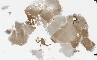
Oligodendroglioma is a diffusely infiltrating glioma that by definition harbours mutations in isocitrate dehydrogenase (IDH1 or IDH2) and codeletion of chromosomal arms of 1p and 19q. They are classified as WHO grade II. Typically, they occur in younger adults and preferentially arise in the cerebral hemispheres. Microscopically, oligodendroglioma is composed classically of uniform round cells with perinuclear haloes along with microcalcifications, mucoid/cystic degeneration, and delicate branching (“chicken-wire”) vasculature. The presence of brisk mitotic activity, microvascular proliferation, and necrosis correspond to anaplastic oligodendroglioma, WHO grade III. Note that the diagnosis of oligodendroglioma requires demonstration of IDH1/2 mutation (typically by IDH1 R132H immunohistochemistry – this slide) and 1p/19q-codeletion (e.g. by FISH, positive in this case). 1p/19q-codeletion is mutually exclusive with ATRX or p53 mutations seen in IDH-mutant astrocytomas. Median survival is in the vicinity of 10 to 20 years.
Gao, A., Munoz, D. Brain, Oligodendroglioma, IDH1 R132H stain. Digital Laboratory Medicine Library, Dept of Laboratory Medicine & Pathobiology, University of Toronto. Published
. Accessed December 17, 2025. https://dev.dlml.cflabs.ca/image/brain-oligodendroglioma-idh1-r132h-stain-lmp80864