Paget disease is a rare form of breast cancer, presenting as skin changes.
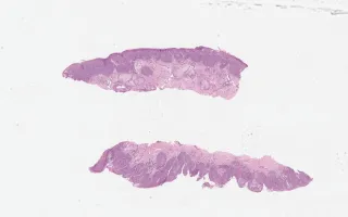
The malignant epithelial cells are seen in the epidermis of nipple/areola. Differential diagnoses to consider include melanoma, Toker cell hyperplasia and Bowen’s disease. One can appreciate the sparing of the basal layer, which is helpful in distinguishing from squamous cell carcinoma. The combination is immunostains—HMWK, CK7-positivity, with HER2-positivity—is helpful. One can appreciate different staining intensity with HMWK, with the intensity being much higher in the keratinocytes. CAM5.2 is expected to be negative (vs. positive in Toker cells). HMB45 can be used to distinguish with melanoma.
This slide shows H&E stain. See related content for HMWK, CK7, and HER2 stains.
See LMP17249-Breast, Paget disease of the nipple, for another example of this entity (link in related content).
Yoon, J., Bocicariu, A. Breast, Paget disease, H&E stain. Digital Laboratory Medicine Library, Dept of Laboratory Medicine & Pathobiology, University of Toronto. Published
. Accessed December 17, 2025. https://dev.dlml.cflabs.ca/image/breast-paget-disease-he-stain-lmp71115