Details
Patient presented with acute appendicitis and perforation. Appendectomy was performed (see Related Content), and the patient was followed. A subsequent recurrent of disease occurred necessitating surgical resection of the colon (this specimen).
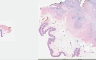
This case represents a goblet-cell carcinoid that has recurred and is now invasive within the wall of the colon (this slide). Conventional-type adenocarcinoma has also arisen within the goblet-cell carcinoid ("adenocarcinoma ex goblet-cell carcinoid" or mixed carcinoid-adenocarcinoma). This slide is an Alcian Blue stain highlighting the presence of mucin in tumour cells.
Well-differentiated neuroendocrine tumours ("carcinoids") of the appendix are usually found incidentally at appendectomy. When large or not confined to the tip, they can cause obstructive symptoms and/or appendicitis. Although uncommon, they account for a majority of appendiceal tumours. They are considered tumours of uncertain malignant potential. Goblet-cell carcinoid is a variant that shows both glandular and endocrine differentiation, with cells resembling normal intestinal goblet cells. Goblet-cell carcinoid is more aggressive than conventional carcinoids.
The prior appendiceal tumour was stained for EMA (LMP99001) and Alcian Blue (LMP84148), highlighting the presence of epithelial differentiation and mucin production, respectively. The original H&E slide is also linked (LMP28939) - see Related Content.
Four years later, the patient developed recurrent disease in which a malignant component was identified. A right hemicolectomy was performed, showing both a goblet-cell carcinoid (LMP26556) and invasive adenocarcinoma (LMP24767) component - see Related Content.