Details
Known Stage IIIA, grade II Follicular lymphoma, on active surveillance; new left tonsillar mass.
Follicular lymphoma (FL) accounts for about 20% of all non-Hodgkin lymphomas. It mainly involves the lymph nodes, but involvement of spleen, bone marrow, Waldeyer ring, skin, duodenum, ocular adnexa, breast and testis can also occur.
Histologically, it shows compact neoplastic follicles composed of germinal centre B-cells (both centrocytes and centroblast) surrounded by attenuated or sometimes absent mantle zones.
FL is graded by counting the absolute number of centroblasts / HPF in 10 neoplastic follicles. Three grades are assigned as follows: grade 1 = 0-5 centroblasts/HPF, grade 2 = 6-15 centroblasts/HPF, and grade 3 > 15 centroblasts/HPF. Grades 1 and 2 are both clinically indolent, and as such a distinction between the two is not necessary. Grade 3 is split into 3A (centrocytes still present) as in our case, and 3B (composed entirely of centroblasts).
Immunohistochemically, the neoplastic cells express B-cell associated antigens (CD19, CD20, CD22, CD79a) and are BCL-2+, BCL-6+and CD10+. Some grade 3B lymphomas may lose CD10 expression.
Cytogenetically, FL is characterized by the t(14;18)(q32;q21) translocation and BCL2 gene rearrangements.
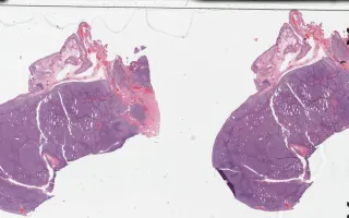
Sections in this case show effacement of the lymph node architecture by an abnormal lymphoid proliferation with nodular pattern. There is an average of more than 15 centroblasts per 40x high power field amongst centrocytes. The follicular architecture is maintained throughout. No sheeting or clusters of centroblasts are identified.
Immunohistochemistry studies demonstrated that the neoplastic cells were positive for CD20, CD10, BCL-2 and BCL-6 (see related content). They were negative for CD3, CD5, CD23, CD43 and cyclin D1 (not shown). CD21 revealed the presence of a follicular dendritic cell meshwork (not shown).
Flow cytometry analysis revealed a population of B-cells that were positive for CD19 and CD20 with co-expression of CD10 and clonal kappa light chain restriction. Overall, the combined morphologic and immunophenotypic features were consistent with a follicular lymphoma, grade 3A.
This slide shows H&E stain. See Related Content for CD20, CD10, BCL6, and BCL2 stains.