The cell of origin for mantle cell lymphoma (MCL) appears to be naïve pre-germinal centre B cells (as suggested by unmutated VH region genes), present in primary & secondary follicles (mantle zone). Architectural effacement by small-to-medium-sized, angulated cells can result in nodular, diffuse or mantle-zone growth patterns.
Much like chronic lymphocytic leukaemia (CLL), MCL is a B cell neoplasm (i.e. CD19, CD20, CD79B, PAX5-positive) with CD5 expression. However, unlike typical CLL, MCL is CD23-negative (although rare cases can have dim CD23 expression). CD20 is generally brighter than for CLL on flow cytometry, which can further the confusion between CLL and MCL. This case was MUM1-positive, which can be seen in ~35% of MCLs. In contrast, benign mantle cells are MUM1-negative. Both benign mantle and MCL cells are generally negative for the germinal centre markers CD10 and BCL6.
Unlike most CLL cases, MCL is a clinically aggressive disease, associated with t(11;14)(q13;q32) that results in Cyclin D1 overexpression. FISH can examine for the CCND1-IgH fusion gene.
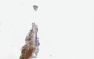
This slide shows Cyclin D1 stain, H&E stain and immunohistochemistry for CD20, CD5, BCL2, and MUM1 are included in the Related Content section.
Also see LMP70330 - Lymph Nodes, Mantle cell lymphoma (link in Related Content).
Yoon, J., Yan, J. Lymph Nodes, Mantle cell lymphoma, cyclin D1 stain. Digital Laboratory Medicine Library, Dept of Laboratory Medicine & Pathobiology, University of Toronto. Published
. Accessed December 17, 2025. https://dev.dlml.cflabs.ca/image/lymph-nodes-mantle-cell-lymphoma-cyclin-d1-stain-lmp65451