2 year old male, presented with nasal polyps and multiple bone lesions.
Case Discussion
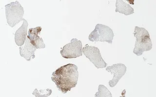
Langerhan cell histiocytosis (LCH) is a rare histiocytic disorder, believed to represent a clonal proliferation of Langerhan cells. It has been historically referred to as histiocytosis X, Hand-Schuller-Christian disease and Letterer-Siwe disease. The multisystem/multifocal form predominantly affects children with a male predominance. It commonly presents with lytic bone lesion(s). However, it can also involve skin, liver, spleen, lymph node, and bone marrow. Microscopically, the neoplastic cells have a “coffee-bean” appearance with elongated nuclei and nuclear grooves, along with abundant eosinophilic cytoplasm. The Langerhan cells are embedded in a milieu composed of variable number of eosinophils, neutrophils and small lymphocytes. These cells express CD1a, langerin (CD207), and S100 protein. In this case, within the fragments of the nasal polyp resection, there are numerous Langerhan cells and eosinophils within a loose, edematous inflammatory mucosa.
This slide shows CD1a stain. See related content for H&E and S100 stains.
Lou, S., Musani, R. Nasal Cavity, Langerhan cell histiocytosis, CD1a stain. Digital Laboratory Medicine Library, Dept of Laboratory Medicine & Pathobiology, University of Toronto. Published
. Accessed December 17, 2025. https://dev.dlml.cflabs.ca/image/nasal-cavity-langerhan-cell-histiocytosis-cd1a-stain-lmp88726