Case Discussion
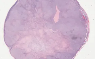
The ovarian stroma is a specialized supportive tissue deriving from the gonadal ridge (mesoderm). Tumours composed of more plump spindle cells containing small lipid droplets are classified as thecomas. Thecomas can be difficult to distinguish from the solid pattern of adult‐type granulosa tumour (considered a low‐grade malignancy). Both thecomas and granulosa cells are positive for calretinin and inhibin (see Supplementary Images in the Related Content section). However, in thecomas, a reticulin silver stain surrounds individual stromal cells. (Link to slide with reticulin stain provided the Related Content section.)