Obstetric ultrasound performed at 15 weeks gestation showed no fetal heart tone. Subsequently, products of conception were delivered intact and sent for pathologic examination.
Case Discussion
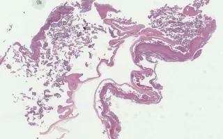
A common cause of spontaneous abortion and stillbirth is fetal chromosomal abnormality, especially trisomy of a somatic chromosome (especially 13, 16, 18, 21). Trisomy can also result in abnormalities in a placenta that resemble the changes seen with molar pregnancy. The villous changes seen in trisomy can sometimes be distinguished from a partial mole by the lack of central cavitation and the presence of abundant spindled stromal cells within the villous tissue. Examination of the fetus can be useful in establishing the diagnosis. In this particular case, there was a stillborn fetus with characteristics suggestive of Trisomy 18 (cleft palate, omphalocele, abnormalities of the digits) that prompted karyotyping of fresh tissue. When fresh tissue is not available, testing that can be done on paraffin blocks include ploidy analysis and FISH. More sensitive and specific is tissue genotyping where maternal tissue is compared to fetal tissue with respect to a wide range of short tandem repeats within the somatic and sex chromosomes.
Parra-Herran, C., Chang, M. Placenta, non‐molar trisomy. Digital Laboratory Medicine Library, Dept of Laboratory Medicine & Pathobiology, University of Toronto. Published
. Accessed December 17, 2025. https://dev.dlml.cflabs.ca/image/placenta-non%25E2%2580%2590molar-trisomy-lmp22150