Details
The patient presented with a left-sided verrucous plaque in the groin. He had multiple lesions on both the left and right side of the scrotum, and along the lower shaft of the penis that first appeared a year and a half prior to presentation. The right sided lesions and the lesions along the shaft of the penis resolved. However, the left sided lesion persisted. The patient's past medical history is significant for HIV, Hepatitis B and hypertension. He was on antiretroviral therapy and his viral load was undetectable.
Cutaneous fungal infections can be primary or secondary. Primary lesions arise from direct inoculation and secondary lesions arise from haematogenous dissemination. Immunocompromised patients are at increased risk for such infections. HIV patients are at increased risk for invasion when CD4 count is under 50 mm^3. The most common opportunistic fungal organism is Candida. Patients usually present with plaques, papules, bullae, abscesses, pustular lesions or subcutaneous granulomas.
On microscopy, this case shows granulomas composed of tightly packed mononuclear cells. Granulomas can range from a well demarcated process to a diffuse inflammatory cell infiltrate with loose granulomas. Necrotizing granulomas have a central focus of neutrophils which is suggestive of an infectious process. In this case, molecular testing (by PCR) confirmed infection by Candida albicans.
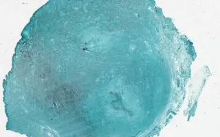
Special staining should include a Periodic Acid-Schiff (PAS) stain and a Grocott methenamine silver (GMS) stain which, in the case of a fungal infection will demonstrate fungal hyphae. A stain for acid fast bacilli (AFB) should also be performed to rule out mycobacterial infection.
This slide shows GMS stain. See Related Content for H&E stain.