32 year-old woman with a history of a long-standing pigmented lesion on her buttocks. The lesion increased in size during pregnancy and was subsequently biopsied.
Case Discussion
Cutaneous malignant melanoma is a neoplasm that arises from pigment producing cells, melanocytes. It is primarily caused by exposure to UV radiation.
The incisional biopsy demonstrates a multinodular proliferation of densely pigmented melanocytes within the dermis. The bleached H&E slide reveals nuclear atypia and 2 mitoses per mm². The overlying superficial dermis and epidermis are uninvolved. This lesion likely represents a primary dermal melanoma with a blue nevus-like morphology. Alternatively, it may represent a melanoma in which the superficial and radial growth patterns have not been sampled.
It is important to note that the Breslow thickness may not be representative of tumour stage given lack of surface epithelium involvement.
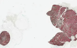
The immunohistochemistry shows lesional cells to be positive for HMB45 and Melan-A.
This slide shows Melan-A stain, see related content for H&E, HMB45, and bleached H&E stains.
Mrkonjic, M., Sade, S. Skin, Malignant melanoma, Melan-A stain. Digital Laboratory Medicine Library, Dept of Laboratory Medicine & Pathobiology, University of Toronto. Published
. Accessed December 17, 2025. https://dev.dlml.cflabs.ca/image/skin-malignant-melanoma-melan-stain-lmp25475