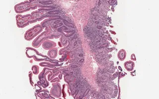
Autoimmune enteropathy is a rare cause of intractable diarrhea. The childhood form is often associated with IPEX (immunodysregulation, polyendocrinopathy, enteropathy, X-linked) syndrome and APECED (autoimmune polyendocrinopathy, mucocutaneous candidiasis, and ectodermal dystrophy) syndrome. The typical histological findings are villous atrophy, an increase in lamina propria inflammation, an increase in intraepithelial lymphocytes (IELs) (primarily at the base of crypts), an increase in apoptotic bodies, and occasionally a lack of goblet cells, Paneth cells and/or endocrine cells. In small intestinal biopsy, note the absence of goblet cells and Paneth cells. The villous architecture here was preserved with no IELs. In the large intestine, there was absence of goblet cells, active inflammation with increased basal crypt IELs and prominent basal crypt apoptoses. The FOXP3 immunostain shows the presence of T-regulatory cells thus excluding IPEX syndrome. The presence of enteroendocrine cells was confirmed by synaptophysin and chromogranin which helped to rule out a stem-cell related lack of development.
This slide shows H&E stain of the small intestine. See Related Content for H&E stain of large intestine and FOXP3 stain.
Forse, C., Siddiqui, I. Small Intestine, Autoimmune enteropathy. Digital Laboratory Medicine Library, Dept of Laboratory Medicine & Pathobiology, University of Toronto. Published
. Accessed December 17, 2025. https://dev.dlml.cflabs.ca/image/small-intestine-autoimmune-enteropathy-lmp75633