Trisomy 21, history of bilateral cryptorchidism, right testicular mass.
Case Discussion
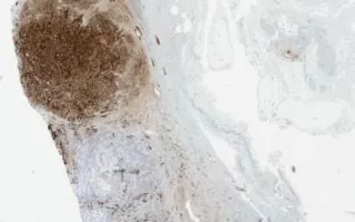
Yolk sac tumours are the most common testicular tumours in infants and young children. They may also occur in adults, usually as a component of a mixed germ cell tumour. Microscopically, yolk sac tumours often have a heterogenous appearance with multiple architectural patterns. This case demonstrates a mixture of microcystic, reticular, glandular/tubular, and solid patterns amongst a variably myxoid stroma. Focal areas consistent with Schiller-Duval bodies are also seen (fibrovascular cores with tumour cells, surrounded by cystic spaces and a second layer of tumour cells, resembling glomeruli). Intercellular eosinophilic globules are also seen throughout the tumour and are composed of alpha-fetoprotein (AFP) and alpha-1-antitrypsin. AFP staining by immunohistochemistry is characteristic of this tumour as seen in this slide. See Related Content section for H&E stain.
Yang, H., Chami, R. Testis (undescended), Yolk-sac tumour, AFP stain. Digital Laboratory Medicine Library, Dept of Laboratory Medicine & Pathobiology, University of Toronto. Published
. Accessed December 17, 2025. https://dev.dlml.cflabs.ca/image/testis-undescended-yolk-sac-tumour-afp-stain-lmp67764