18-year-old woman originally from Central America with recent onset of headaches. Neuroimaging shows a cerebellar cystic lesion with enhancing mural nodule.
Case Discussion
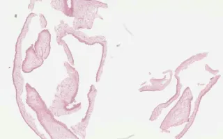
Neurocysticercosis is a CNS infection caused by the larval form of Taenia solium (pork tapeworm). This occurs due to accidental ingestion of adult tapeworm eggs, which are shed in feces. The eggs eventually hatch, enter the bloodstream, implant in tissues (typically brain, skeletal muscle, or eye), and develop into the larval form (cysticercus). The most common clinical presentation is seizures. Microscopically, the cyst wall has 3 layers: an outer eosinophilic cuticular layer, a middle cellular layer, and an inner reticular layer. Treatment can be surgical to relieve CSF obstruction and/or medical with antiparasitic drugs.
This slide is taken from a cyst wall. See Related Content for section from a T. solium larva.
Gao, A., Kiehl, TR. Brain, Neurocysticercosis. Digital Laboratory Medicine Library, Dept of Laboratory Medicine & Pathobiology, University of Toronto. Published
. Accessed December 17, 2025. https://dev.dlml.cflabs.ca/image/brain-neurocysticercosis-lmp33454