Mantle cell lymphoma (MCL) accounts for approximately 3-10% of non-Hodgkin lymphomas. It mainly involves the lymph nodes as well as spleen and bone marrow. Extra nodal sites include GI and Waldeyer ring. Of note, MCL is the most common cause of multiple lymphomatous polyposis.
Histologically, MCL is composed of monomorphic, small to medium-sized cells with irregular nuclear membranes, arranged in a nodular, diffuse, or follicular configuration. Many morphological variants are recognized with blastoid and pleomorphic variants having the worst prognosis.
Immunohistochemically, the neoplastic cells express BCL-2, cyclinD1, CD5, FMC-7 and CD43, but are negative for CD23, CD10 and BCl-6. Rare cases of CD23 positivity have been reported.
Cytogenetically, MCL is characterized by the t(11;14)(q13;q32) translocation between IgH and cyclin D1 (CCND1) genes. In rare cases, the t (11;14) translocation and cyclinD1 expression are lacking. Instead, these cases express cyclinD2, cyclinD3 or SOX11.
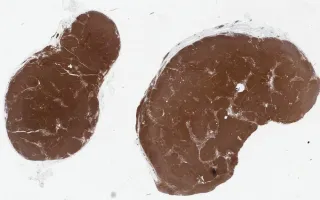
This slide shows CD5 stain. See Related Content for H&E, CD23 and cyclin D1 stains.
Starova, B., Ghorab, Z. Lymph Nodes, Mantle cell lymphoma, CD5 stain. Digital Laboratory Medicine Library, Dept of Laboratory Medicine & Pathobiology, University of Toronto. Published
. Accessed December 17, 2025. https://dev.dlml.cflabs.ca/image/lymph-nodes-mantle-cell-lymphoma-cd5-stain-lmp95959