Chronic minimally active gastritis with H. heilmannii
Clinical History
Inflammation of the esophagus.
Case Discussion
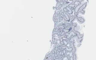
The gastric cardia can extend into the lower esophagus, and become inflamed causing symptoms of esophagitis. Sections show gastric-type mucosa with preserved glandular architecture. There is a mild increase in lymphoplasmacytic infiltrate in the superficial lamina propria which is more prominent in antral biopsy (part C). There is minimal focal active inflammation with single neutrophils infiltrating the pits in the body biopsy (part D). There are scattered "corkscrew" shaped coiled spirals of Helicobacter heilmannii organisms present on the luminal surface as well as coccoid forms infiltrating the crypt epithelium, seen both on H&E (see Related Content) and immunostains (this slide). There are no granulomas or giant cells.
Peerani, R., Siddiqui, I. Gastroesophageal junction, Helicobacter heilmannii gastritis, Helicobacter stain. Digital Laboratory Medicine Library, Dept of Laboratory Medicine & Pathobiology, University of Toronto. Published
. Accessed December 17, 2025. https://dev.dlml.cflabs.ca/image/gastroesophageal-junction-helicobacter-heilmannii-gastritis-helicobacter-stain-lmp55820