18 year old female with a history of tuberous sclerosis, who presented with blood per rectum and a long history of intestinal pseudo-obstruction. MRI showed asymmetric thickening of the rectum. The patient underwent a proctectomy.
Case Discussion
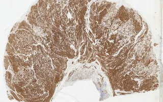
Patients with tuberous sclerosis complex (TSC) are predisposed to a number of tumours, including pulmonary lymphangioleiomyomatosis (LAM) and renal angiomyolipomas (AML). This case is a rare example of leiomyomatosis-like lymphangioleiomyomatosis involving the rectum. Microscopically, the rectal wall is thickened with ill-defined nodules of spindle cells and vessels. LAM and AML are both part of the spectrum of perivascular epithelioid tumours (PEComas). Immunohistochemical stains showed that the lesional cells were diffusely positive for desmin and SMA, and had patchy HMB-45, ER, and PR positivity. This resection specimen also contained an incidental well-differentiated neuroendocrine tumour.
This slide shows H&E stain, see Related Content for HMB-45 and SMA stains.
Kolin, D., Somers, G. Rectum, lymphangio-leiomyomatosis, SMA stain. Digital Laboratory Medicine Library, Dept of Laboratory Medicine & Pathobiology, University of Toronto. Published
. Accessed December 17, 2025. https://dev.dlml.cflabs.ca/image/rectum-lymphangio-leiomyomatosis-sma-stain-lmp44369