57 year-old woman with intra-abdominal lymphadenopathy; mesenteric lymph node biopsy was performed.
Case Discussion
Follicular lymphoma is a B-cell neoplasm composed of germinal center B cells (centrocytes and centroblasts). It makes up about 20% of non-Hodgkin lymphoma in USA and Western Europe. Patients are usually asymptomatic (even when disease is disseminated) at presentation with an overall 10-year survival of up to 80%. It is characterized by closely packed neoplastic follicles, uniform in size and shape. The neoplastic follicles contain variable amounts of centrocytes and large centroblasts. Grading is based on the number of centroblasts per high power field and is associated with prognostic and therapeutic significance.
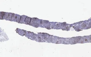
The needle core biopsy shows nodular proliferation of mostly small cleaved atypical lymphoid cells (centrocytes) and less numerous large non-cleaved atypical lymphoid cells (centroblasts,
Shao, TS., Yan, J. Lymph Nodes, Follicular lymphoma, MIB1 stain. Digital Laboratory Medicine Library, Dept of Laboratory Medicine & Pathobiology, University of Toronto. Published
. Accessed December 17, 2025. https://dev.dlml.cflabs.ca/image/lymph-nodes-follicular-lymphoma-mib1-stain-lmp85187