Biopsy of ulcerated leg lesion; also has a history of pneumonia, unresponsive to antibiotics.
Case Discussion
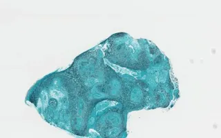
Blastomyces dermatitidis is a fungal organism which can cause pneumonia and skin infections. The H&E image (see Related Content) shows epidermal/superficial dermal microabscesses with neutrophils as well as broad based, thick walled blastomyces organisms. The GMS silver stain (this slide) highlights the organisms (in black).
Blastomyces are found in the soil in the Ohio River and Mississippi River deltas, and the Great Lakes-St. Lawrence region. It commonly presents as pneumonia or as a skin infection. The cutaneous presentation often results in verrucous/papillary or ulcerative lesions. Histologically, blastomyces spores are typically 8-15 microns in size and exhibit broad-based budding. They can often be found in dermal microabscesses. The differential diagnosis would include other infections, including other fungi, but blastmycosis can mimic malignancy such as squamous cell carcinoma due to the presence of squamous hyperplasia.
Wang, T., Chung, C. Skin, Cutaneous Blastomycosis, GMS stain. Digital Laboratory Medicine Library, Dept of Laboratory Medicine & Pathobiology, University of Toronto. Published
. Accessed December 17, 2025. https://dev.dlml.cflabs.ca/image/skin-cutaneous-blastomycosis-gms-stain-lmp21539