Granular cell tumours are benign tumours which may occur at virtually any anatomical site and across a wide range of ages. The most common anatomical site is the tongue. They are said to be likely neuroectodermal in nature and may arise from Schwann cells.
In the breast, this tumour can radiologically and clinically mimic a carcinoma. On gross examination, the borders can be irregular and the tumour is firm and tan/yellow in colour. Microscopically, these tumours are composed of large polygonal cells with eosinophilic to gray granular cytoplasm with small, centrally located bland nuclei. Atypia and mitotic activity are generally absent though may be scattered. The granular quality of the cytoplasm is attributed to an accumulation of lysosomes.
These lesions are typically treated with complete excision. The local recurrence rate in these benign lesions is quite low, and recurrence usually reflects incomplete excision.
Malignant granular cell tumour is rare (only 2-3% of all granular cell tumours) and is most often seen in the deep soft tissue of adults. According to Fletcher, any deep seated or large lesion with pleomorphism, necrosis, and frequent mitoses should be regarded with suspicion (Diagnostic Histopathology of Tumors, 3rd ed, 2007, p. 1747).
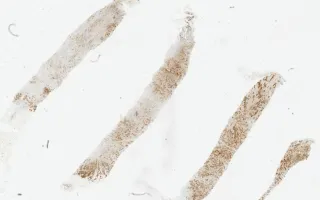
This slide shows CD68 stain. The patient's biopsy and excision material is shown in the Related Content section. S100 immunostain is also included. The tumour was negative for pankeratin.
Hodgson, A., Lu, F. Breast, Granular cell tumour, CD68 stain. Digital Laboratory Medicine Library, Dept of Laboratory Medicine & Pathobiology, University of Toronto. Published
. Accessed December 17, 2025. https://dev.dlml.cflabs.ca/image/breast-granular-cell-tumour-cd68-stain-lmp82734