Meningiomas are tumours that arise from meningothelial cells of the arachnoid. Most meningiomas are benign (WHO grade I). The WHO classification of tumours describe nine histologic variants of grade I meningiomas which may mimic other tumours.
This case is an angiomatous variant of meningioma which demonstrates a highly vascular and cellular tumor composed of round to oval cells with a syncytial growth pattern. The cells have numerous pseudonuclear inclusions and visible nucleoli. The cells are distributed amongst a richly vascular network of large dilated branching vessels and innumerable small vessels. Degenerative phenomena such as nuclear pleomorphism and vascular hyalinization may also be seen.
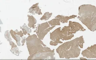
Immunohistochemistry is supportive of meningeal origin with positivity for EMA, PR, E-cadherin, and D2-40. CD34 highlights an extensive vascular network of small vessels but the tumour cells are negative.
This slide shows D2-40 stain. See Related Content for H&E, CD34, EMA, PR, and E-cadherin stains.
Yang, H., Keith, J. Brain, Angiomatous meningioma, D2-40 stain. Digital Laboratory Medicine Library, Dept of Laboratory Medicine & Pathobiology, University of Toronto. Published
. Accessed December 17, 2025. https://dev.dlml.cflabs.ca/image/brain-angiomatous-meningioma-d2-40-stain-lmp76640