Skin biopsy of left medial leg in an 86 year-old male on Pembrolizumab for melanoma.
Case Discussion
Desmoplastic melanoma typically presents as an amelanotic lesion within sun-exposed areas and often resembles scar tissue. Histologically, the lesion is often ill-defined with atypical spindled cell melanocytes infiltrating the dermis. Overlying lentigo maligna and lymphoid aggregates may be seen. Neurotropism is also a common finding. Immunohistochemical staining typically shows S100 and SOX10 positivity, while HMB-45 is negative.
In this case, the biopsy demonstrates a subtle case of desmoplastic melanoma with scattered malignant cells between collagen bundles. Neurotropism is also seen.
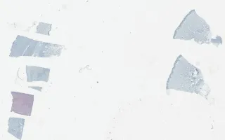
SOX10 and S100 stains highlight malignant spindled cells infiltrating the dermis.
This slide shows SOX10 stain (using a red chromogen). See related content for H&E and S100 stains.
Pun, C., Sade, S. Skin, Desmoplastic melanoma, SOX10 stain. Digital Laboratory Medicine Library, Dept of Laboratory Medicine & Pathobiology, University of Toronto. Published
. Accessed December 17, 2025. https://dev.dlml.cflabs.ca/image/skin-desmoplastic-melanoma-sox10-stain-lmp55056