Hydatid disease is a zoonosis caused by the larval stages of the parasitic tapeworm of the genus Ecchinococcus. The right lobe of the liver is the most commonly involved site; however the infectious agent may also involve the lungs, kidneys, spleen, brain and muscoskeletal system.
The outer chitinous (or fibrous laminar) layer is the key histologic feature. Clinicians are often aware of the diagnosis before the report, based on the serology. The PAIR sequence (puncture, aspiration, injection, re-aspiration) is the commonly used management strategy that allows for the direct diagnosis.
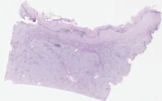
This slide shows trichrome stain. See related content for H&E stain.
Yoon, J., Fischer, S. Liver, Hydatid disease, trichrome stain. Digital Laboratory Medicine Library, Dept of Laboratory Medicine & Pathobiology, University of Toronto. Published
. Accessed December 17, 2025. https://dev.dlml.cflabs.ca/image/liver-hydatid-disease-trichrome-stain-lmp82890