Hysterectomy for fibroids, post fibristal treatment.
Case Discussion
Highly cellular leiomyoma is a benign leiomyoma variant. It is an important entity because it mimics the appearance of an endometrial stromal neoplasm. The distinguishing features of highly cellular leiomyoma are its very low mitotic activity (and MIB1 expression), fascicular growth, and the diffuse presence of the thick walled vessels characteristic of smooth muscle tumours. Moreover, the highly cellular variant is often negative for the usual markers of the smooth muscle differentiation and shows CD10 reactivity. As such, these stains are of reduced diagnostic utility.
The findings in this case are of a 9.5 cm intramural mass with a rounded shape and some irregularities on the border in which cellular areas merge with the surrounding myometrium. The cells are bland spindle cells with scant cytoplasm arranged in diffuse sheets and fascicles. The tumour forms abundant cleft like spaces and thick walled vessels are present. Mitoses number fewer than 1 per 10 HPFs. Immunohistochemistry performed at the referring institution show that the tumour cells are positive for beta catenin, myosin, CD10, and negative for desmin. Both ER and PR stains are strongly diffusely positive within the tumour cells. Based on these findings this tumour is best classified as a highly cellular leiomyoma.
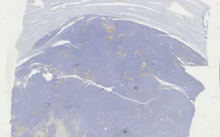
This slide shows MIB1 stain. See related content for H&E, CD10, caldesmon, and desmin stains.
Parvinnejad, N., Chang, M. Uterus, Highly cellular leiomyoma, MIB1 stain. Digital Laboratory Medicine Library, Dept of Laboratory Medicine & Pathobiology, University of Toronto. Published
. Accessed December 17, 2025. https://dev.dlml.cflabs.ca/image/uterus-highly-cellular-leiomyoma-mib1-stain-lmp27803