Details
Large peritoneal mass.
Anaplastic lymphoma kinase negative (ALK -) anaplastic large cell lymphoma (ALCL) is a T-cell neoplasm occurring in adults, with a slight male preponderance. They are relatively rare in that they represent only 2-3% of all non-Hodgkin lymphoma, and approximately 10% of all T-cell lymphomas.
These tumours are most commonly composed of sheets of large and pleomorphic cells, often with prominent nucleoli. When involving a lymph node, a sinusoidal pattern of infiltration can also occur. "Hallmark cells", the prototypical cell type in ALK+ ALCL with a horseshoe-shaped or donut-shaped nucleus and eosinophilic paranuclear region, are less commonly seen in ALK- tumours. CD30 staining is strong and uniform, and aberrant T-cell immunophenotypes are common. Frequent expression of cytotoxic molecules is also a feature. As the name implies, ALK expression is not detected. The morphological differential diagnosis includes ALK+ ALCL, peripheral T cell lymphoma NOS, primary cutaneous ALCL (when confined to cutaneous sites) and diffuse large B cell lymphoma. When features such as sclerosis and eosinophils are seen, Hodgkin lymphoma is also considered.
At least 3 molecular subsets have been identified: 6p25.3/DUSP22 translocation in 30% (good prognosis), 3q28/TP63 in ~ 8 % of cases (poor prognosis), and the remaining cases are classified as double negative (intermediate prognosis).
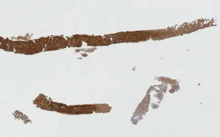
In this case, cores of tissue are seen to be infiltrated by sheets of large pleomorphic cells with brisk mitotic activity and evident nucleoli. There is necrosis and small vessels are seen scattered throughout the sheets of tumour cells. The tumour cells are positive for: CD45, CD30 (diffuse), CD7 (decreased), CD5 (partial), CD4 (partial), EMA (partial), TIA1, and MUM1; and negative for: CD2, CD3, CD8, and ALK1 (see Related Content for selected IHC stained slides). Granzyme B IHC was also attempted in this case to further support T cell origin (cytotoxic marker), however technical issues precluded testing.
Concurrent flow cytometry was non-contributory in this case.
This slide shows CD30 stain. See related content for H&E and ALK1 stains.
1) WHO Classification of Tumours of Haematopoietic and Lymphoid Tissues. World Health Organization Classification of Tumours. Lyon, France: IARC; 2008