64-year-old female with left axillary lymphadenopathy.
Case Discussion
Angioimmunoblastic T-cell Lymphoma is a neoplasm of mature T follicular helper (TFH) cells. It is seen mainly in older male adults and accounts for up to one third of non-cutaneous T-cell lymphomas.
It is clinically aggressive and often presents at a later stage with generalized lymphadenopathy. Systemic symptoms including polyclonal hypergammaglobulinaemia, skin rash, pruritus, pleural effusion, arthritis, and ascites are also common.
Affected lymph nodes show partial or total effacement of the architecture with a prominent proliferation of high endothelial venules (HEVs). Small clusters of the neoplastic cells are often seen adjacent to the HEVs. These cells are small to medium lymphocytes with clear and pale cytoplasm distinct cell membranes, and minimal atypia. These are seen in a rich reactive background of lymphocytes, histiocytes, plasma cells, and eosinophils.
EBV-positive B cells are nearly always present, and in some cases constitute a significant part of the cellular infiltrate
Tumor cells express most pan-T-cell antigens (e.g. CD3, CD2, and CD5) and, in the vast majority of cases, are positive for CD4. They also express normal TFH cells phenotype: CD10, CXCL13, ICOS, BCL6, and PD1.
T cell receptor genes show clonal rearrangements in 75-90% of cases.
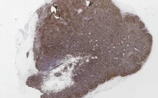
This slide shows CD3 stain. See related content for H&E stain.
Algawahmed, F., Delabie, J. Lymph Nodes, Angioimmunoblastic T-cell Lymphoma, CD3 stain. Digital Laboratory Medicine Library, Dept of Laboratory Medicine & Pathobiology, University of Toronto. Published
. Accessed December 17, 2025. https://dev.dlml.cflabs.ca/image/lymph-nodes-angioimmunoblastic-t-cell-lymphoma-cd3-stain-lmp74984