Left breast circumscribed mass, with radiologic calcifications, slowly growing over 10 years.
Case Discussion
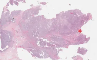
Malignant phyllodes tumours are fibroepithelial neoplasms with malignant stromal growth. Criteria for malignancy included stromal overgrowth, stromal hypercellularity, stromal cell atypia, mitotic activity, and invasive tumour borders. In this case, the diagnosis is complicated because there is no epithelial component and it is therefore difficult to prove this lesion’s fibroepithelial origin. The differential diagnosis is mainly between a primary breast sarcoma and malignant phyllodes tumour. Based on the circumscribed and relatively indolent clinical course, the diagnosis of malignant phyllodes tumour with osteosarcomatous overgrowth was favoured in this case.
The present image highlights the presence of osteoid and osteoclast-like giant cells. See image (LMP16263) in related content which highlights the very circumscribed borders of the lesion.
Chang, M. Breast, Malignant phyllodes tumour, H&E stain. Digital Laboratory Medicine Library, Dept of Laboratory Medicine & Pathobiology, University of Toronto. Published
. Accessed December 17, 2025. https://dev.dlml.cflabs.ca/image/breast-malignant-phyllodes-tumour-he-stain-lmp27319