Right breast carcinoma 14 years ago, treated with lumpectomy, hormonal therapy and radiotherapy. Now presenting with right breast lump and skin bruising. Breast MRI and US show dermal thickening and several masses.
Case Discussion
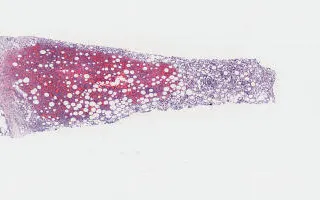
Cosnider secondary angiosarcoma (AS) in patients with prior radiotherapy for breast cancer. Helpful immunohistochemical stains are CD31, CD34, ERG, FVII, and MYC. MYC 8q24 amplification is seen in almost 100% of radiation-induced breast AS. MYC IHC can thus be used to discriminate between secondary AS (MYC+) and radiation-induced lesions (dermatitis, atypical vascular lesion) and primary angiosarcoma which are consistently MYC-negative. Slides provided are H&E (showing cores of adipose tissue infiltrating by sheets of mitotically active, markedly atypical cells with no definitive vessel formation), ERG and MYC IHC both showing diffuse strong nuclear staining.
Morgan, S. Breast, Mammary angiosarcoma, H&E Stain. Digital Laboratory Medicine Library, Dept of Laboratory Medicine & Pathobiology, University of Toronto. Published
. Accessed December 17, 2025. https://dev.dlml.cflabs.ca/image/breast-mammary-angiosarcoma-he-stain-lmp65402