Patient presented with acute appendicitis and perforation. An appendectomy was performed (this specimen), and the patient was followed. A subsequent recurrence of disease occurred (see Related Content).
Case Discussion
Well-differentiated neuroendocrine tumours (“carcinoids”) of the appendix are usually found incidentally at appendectomy. When large or not confined to the tip, they can cause obstructive symptoms and/or appendicitis. Although uncommon, they account for a majority of appendiceal tumours. They are considered tumours of uncertain malignant potential. Goblet-cell carcinoid is a variant that shows both glandular and endocrine differentiation, with cells resembling normal intestinal goblet cells. Goblet-cell carcinoid is more aggressive than conventional carcinoids.
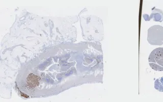
This EMA stain highlights the presence of epithelial differentiation. This appendiceal tumour was also stained for Alcian Blue (LMP63103), highlighting mucin production. The H&E stain is also linked (LMP28939) - see Related Content.
Four years later, the patient developed recurrent disease in which a malignant component was identified. A right hemicolectomy was performed (Alcian Blue: LMP84148, H&E: LMP24767 and LMP26566) - see Related Content.
Mrkonjic, M., Chang, M. Appendix, goblet-cell carcinoid, EMA stain. Digital Laboratory Medicine Library, Dept of Laboratory Medicine & Pathobiology, University of Toronto. Published
. Accessed December 17, 2025. https://dev.dlml.cflabs.ca/image/appendix-goblet-cell-carcinoid-ema-stain-lmp99001