Details
Hematuria, renal mass with invasion of large and small bowel.
Urothelial carcinoma of the upper urinary tract (ureter, pelvis) is relatively uncommon and accounts for approximately 5-10% of all urinary tract carcinomas. It is more common in males compared to females. The risk factors mirror those of the lower urinary tract carcinomas - specifically cigarette smoking, industrial carcinogens, chronic irritation, and phenacetin abuse. Certain genetic factors also play a role - patients with Lynch syndrome have a significant increased risk for upper tract urothelial carcinoma because of defective DNA mismatch repair and microsatellite instability.
Clinically, these tumours can present with flank pain, hematuria or general malaise and weight loss. Microscopically, upper tract urothelial tumours are classified along the same morphologic spectrum as bladder tumours.
In this case, this patient presented with hematuria and was subsequently found to have a right renal mass. On imaging, the tumour was approximately 5cm in its largest dimension, and described as being 'muddy.' There was questionable involvement of both the large and small bowel. Urine cytology had been negative. The clinical differential was between urothelial carcinoma versus renal cell carcinoma.
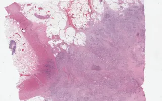
In this slide, a representative section shows a high grade urothelial carcinoma infiltrating the muscularis of the large bowel.
A representative section showing invasion into the musularis of the small bowel is included in the Related Content section.
Upper urinary tract carcinomas are staged as follows:
Ta: Noninvasive papillary urothelial carcinoma
Tis: Urothelial carcinoma in situ
T1: Invasion of lamina propria
T2: Invasion of muscularis propria
T3: Invasion of peripelvic adipose tissue or renal parenchyma (for pelvis) or invasion of periureteral adipose tissue (for ureter)
T4: Invasion of adjacent organs other than the kidney or invasion through the kidney into perinephric adipose tissue.
This case was staged at pT4.
Immunohistochemical stains were done which showed the tumour stained positively for GATA3, CK7, and p63. The microscopic differential was between urothelial carcinoma and collecting duct carcinoma. There was urothelial carcinoma in situ noted in many slides.