This 70-year-old female had a recent history of resected colorectal cancer. She subsequently developed a 3 cm subcapsular liver mass that was initially suspected to be a metastasis. The mass did not respond to adjuvant chemotherapy, so a diagnosis of hepatocellular carcinoma was favoured. She underwent segment V liver resection.
Case Discussion
Clinical features: Hepatic angiomyolipomas are rare tumours, usually solitary and discovered incidentally.
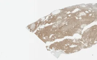
Histological/molecular features: Variable proportions of epithelioid and spindle cells, adipocytes and thick-walled blood vessels. The classic immunohistochemical profile is co-expression of melanocytic markers and smooth muscle markers. Like other PEComas, hepatic angiomyolipomas harbour mutations in the TSC gene.
MacColl, C. Liver, Angiomyolipoma, HMB-45. Digital Laboratory Medicine Library, Dept of Laboratory Medicine & Pathobiology, University of Toronto. Published
. Accessed December 17, 2025. https://dev.dlml.cflabs.ca/image/liver-angiomyolipoma-hmb-45-lmp99237