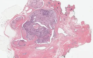
4.6 cm solid intraductal lesion in the retro-areolar area of left breast.
Case Discussion
This fibroepithelial lesion shows an unusual intraductal growth pattern, and mimics a rare entity, intraductal fibroadenomatosis. Intraductal fibroadenomatosis is described as having features of flbroadenomas, papillomas and phyllodes tumours. In this case, the stromal expansion (with focal leaf-like pattern) and stromal cellularity were too pronounced, and the stromal mitotic activity too high (up to 7/10 high power fields), for a diagnosis of fibroadenoma.
Reported cases of intraductal fibroadenomatosis have not featured elevated mitotic activity or stromal atypia (Chung, A.et al., Breast J 2009, 14:193-195; Cummings MC, et al., Virchows Arch 2010, 456:105-106). Although the mitotic activity is more elevated than expected for a benign phyllodes tumour, no other worrisome features are observed - the borders are non-infiltrative, and there is no high-grade cytologic atypia, necrosis, heterologous differentiation or overgrowth in the stromal component. No atypia is identified in the ductal component.
See Related Content for references:
1) Chung, A.et al., Breast J 2009, 14:193-195.
2) Cummings MC, et al., Virchows Arch 2010, 456:105-106
Stephenson, P., Lu, F. Breast, Benign phyllodes tumour. Digital Laboratory Medicine Library, Dept of Laboratory Medicine & Pathobiology, University of Toronto. Published
. Accessed December 17, 2025. https://dev.dlml.cflabs.ca/image/breast-benign-phyllodes-tumour-lmp14365